FORTY HEALTH AND SPORTS ARTICLES.
- Rectal Bleeding
- Abdominal Pain
- Swollen Glands
- Cold Feet
- Chest Pain
- Dry Mouth
- Knee Pains
- Hair Loss
- Breast Discharge
- Frequent Urination
- Heart Attack
- Heart Attack Symptoms
- How to Prevent Heart Attacks
- How to prevent heart attackssportsports equipmentsports equipment storessports fitness equipmentsport books---
- If possible, please also attach 1-2 photo(s) for the equipments/products below jumping stilts Exercise Bikes Elliptical TrainersreadmillsElliptical Trainers Exercise Wheels Chin-Up Bars Home GymsDumbellsExercise ApparelsCycling equipmentsPaintballPaintball equipments--- If possible, please also attach 1-2 photo(s) for the equipments/products below Blood Pressure Monitors Heart Rate Monitors Massage Chairs weight loss products First Aid KitsThermometersPregnancy TestsCholesterol TestsFertility Tests
ARTICLE I
RECTAL BLEEDING - Diagram of the rectum
- Definition And Function Of The Rectum
- Description of Rectal Bleeding
- Sources of Rectal Bleeding
- Colour of the Blood in Rectal bleeding.
- Causes of rectal bleeding.
Diagram of The Rectum
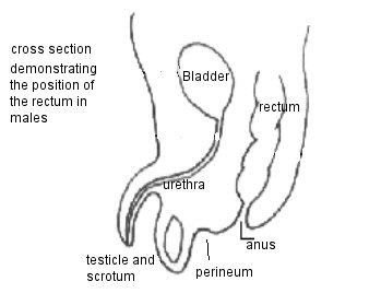
Definition And Function Of The Rectum
The rectum is the posterior end of the digestive tract of the large intestine. The word itself is a misnomer as it comes from the Latin and it originally meant straight, and as the diagram illustrates the human rectum is not. It is a repository for fecal matter, and it has the traverse folds illustrated in the diagram, these folds hold the faeces in position, until you are ready to go to the toilet.
Description of Rectal Bleeding
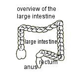
Rectal Bleeding is known medically as hematochezia, it does not indeed refer to bleeding from the rectum but from the anus, which is the orifice to the outside. The blood is bright red but it may be mixed with fecal matter, and or blood clots. The rectum itself lies immediately above the anus. Although the blood may be emanating from the rectum it may also be coming from the intestinal tract, as illustrated in the diagram of the large intestine below. The flow of blood may be mild moderate or severe.
- Mild bleeding is only sufficient to turn the water in the toilet bowl a pale pink. This normally stops on its own accord and does not normally require treatment or hospitalisation.
- Moderate bleeding is heavier and it may be mixed with blood clots in which case the blood is dark, or if it is a bright red colour then it is only fresh blood.
- Severe rectal bleeding is sometimes a single bowel movement that is accompanied by a large blood flow. Or in other cases it is a series of bowel movements accompanied by blood loss
Both moderate and severe cases of rectal bleeding need to be evaluated by a doctor, and in certain cases they require hospitalisation.
Sources of Rectal Bleeding
In the majority of cases rectal bleeding comes from the anus, the rectum, or the colon.
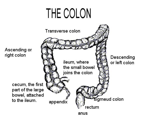
- The Colon
The colon is situated above the rectum, and is part of the digestive tracts. The food passes through the colon once it has left the small intestine. The colon’s job is to remove the water content from the undigested food, which makes it smaller in matter, and then store it until it is eliminated as a stool. The rectum is the last eight inches of the colon and the anal canal or the anus is the orifice, which ejects the stool from the body. Various names, the large intestine, the large bowel, or the lower gastrointestinal tract, refer to the colon, the rectum and the anus, as a whole unit and it comprises of several feet of muscular tubes. Conversely the oesophagus, stomach, duodenum, and small intestine are referred to as the upper gastrointestinal tract
The colon itself is sub divided into three areas the right colon, the transverse colon, and the left colon, as seen on the diagram below.
Colour of the Blood in Rectal bleeding
The colour of the blood is often an indication of the cause of the bleeding. The bleeding can come anywhere from the digestive tract, the higher it comes from the darker it will be. This means in general that if the bleeding is a result of a duodenal ulcer the blood is likely to be darker than if it is coming from the rectum itself where it is more likely to be bright red.
When the right colon bleeds it can contain melena, which are black stools their consistency like tar, accompanied by an objectionable smell. When the blood is in the colon for a longer period of time the blood is broken down into chemicals, which colour and taint the stool. Typically melena occur when the bleeding is coming from higher in the digestive tract such as a duodenal, or stomach ulcer, although occasionally it can be a result of bleeding from the right colon.
However sometimes when these ulcers perforate the blood passes through so quickly it does not have time to break down and it will exit the body as a bright red colour. Evidence of rectal bleeding is not always apparent, when the blood is slight and very slow as is the case with rectal polyps, then the blood loss is absorbed by the stool, and cannot be detected. In these cases a fecal occult blood is necessary to establish whether or not there is blood present.
Causes of rectal bleeding
Haemorrhoids are a collection of tissues within the anus, which contain blood vessels.Contrary to popular belief everyone has them; it is only when they become enlarged or distended that they cause pain and bleed. The resultant blood flow rarely causes anaemia or low blood pressure, but if they are left untreated for years they may cause iron deficiency anaemia.
Anal fissures occur when the lining of the rectum is torn, usually as a result of constipation or a strong bowel movement, though tight anal muscles may cause the problem. It can be an extremely painful condition, because the condition is aggravated by subsequent bowel movements. As it has the same symptoms as haemorrhoids, the light bright blood it is often confused with them, however haemorrhoids do not cause pain during a bowel movement.
Diverticulitis usually develops in people between the age of fifty and sixty years, and the exact cause of their development are not understood. The colon develops little pockets or sacks, and once they have formed they are permanent, the only way of removing them is to remove the affected portion of the colon. This can be done as the diverticula can occur in any part of the colon they are more often observed in the sigmoid colon.
The majority of sufferers will not be aware of the problem until one of these sacks gets an infection or they rupture; it is at this point that the condition becomes diverticulitis. Diverticulitis causes moderate to severe rectal bleeding, it can require a blood transfusion. The colour of the blood is not always an indication, as when the blood is coming from the sigmoid colon it tends to be bright red, as it is when it coming from the right colon if the blood flow is rapid. However there may be melena present in which case it is black, but in general the blood is n the darker side.
The bleeding does tend to stop without any treatment, but often reoccurs, within a short period of time.
Tumours within the colon and rectum originate from the walls of the large intestine, when they are benign they are described as polyps, but when they come malignant they are then called cancer. The amount of blood flow is generally minimal, often bright red, but the darker stools are present which are often maroon and melena may be present. When the blood is mixed with the stool and occurs over a long period of time severe iron deficiency anaemia may become an issue. As many of the cancers of the colon have developed from polyps they are generally surgically removed. Whilst colon cancer is not a rare disease ninety percent of it would be eliminated if we ate the recommended amount of fibre and had regular screening once over the age of fifty. In India where the majority of the people are vegetarians the disease is almost unknown.
A polypectomy is the surgical removal of benign polyps in the colon. This can cause bleeding for weeks after the surgery. However the severity of the bleeding depends on the size of the polyps. The bleeding can be severe and fast flowing and it may be bright red, dark red, maroon, purple or black.
Angiodysplasias is conditions, which generally worsen with age, they are enlarged blood vessels and they are bright red lesions, which radiate like a spider’s web beneath the lining of the colon. They can develop all over the common, but are usually found in the right portion of the colon. The resultant bleeding is not accompanied by pain, but they can cause occult bleeding whereby the blood becomes part of the stool and iron deficiency anaemia can occur. The blood loss can be light or dark or black.
Colitis is defined as inflammation of the colon, whilst proctisis is the equivalent inflammation of the rectum. The inflammation can occur from a plethora of conditions, examples are bacterial or viral infection, ulcerative colitis or proctitis, Crohn’s colitis, ischemic colitis, and radiation colitis or proctitis.
Ulcerative colitis, ulcerative proctitis, and Crohn’s colitis are caused by an overactive immune system. One of the symptoms includes diarrhoea often mixed with blood, but occasionally the rectal bleeding can be moderate to severe, when it originates from ulcerations of the colon.
Infections such as salmonella, shigella, Campylobacter, C. difficile, E. Coli O157:H7, and cytomegalovirus can inflame the colon. They normally only cause diarrhoea with can have traces of blood in it, but in rare occasions moderate to severe bleeding can accompany these infections.
Blood clots can obstruct the arteries that bring blood to the colon, and this happens rapidly causing an inflammation of the colon, which is known as Ischemic colitis. The area affected is normally in the region of the splenic flexure.
Because the blood flow is so immediate it can lead to ulceration within the colon, which is followed, by abdominal cramps, and then rectal bleeding. In these cases the blood flow is minimal and will normally stop within a few days, though the ulcer can take a little longer to heal.
Radiation proctitis follows pelvic radiation occurs after treatment for prostate cancer, this causes permanent changes within the lining of the colon and it may mean that there are instances of rectal bleeding for years. However the level of bleeding is usually mild but if the condition is chronic then anaemia may result.
Meckel’s diverticulum occurs in a very small number of the population from birth. It is a small sack near the junction of the colon and the small intestine. Occasionally they secrete acid that causes small ulcers, and they may bleed. This is the most common cause of rectal bleeding in young adults, teenagers and children. Whilst the flow of blood can be brisk its colour varies from bright red to dark, and often results in purple stools.
When bleeding is both rapid and severe from the upper gastrointestinal tract it is bright red, as it has no time to discolour and is usually a result of stomach or duodenal ulcers. Sometimes a blood vessel ruptures and this causes the blood to leak into the gastrointestinal tract when this occurs it is eliminated from the body via the anus. This event may occur when an aortic graft is being performed to repair an aortic aneurysm. Very rarely rectal bleeding may result from tumours within the small intestine or the rectum.
ARTICLE 2
Abdominal Pain.
In this article:
- Definition of Abdominal pain.
- Causes of abdominal pain
- The four abdominal quadrant
- When to seek medical assistance
- Diagnosis of abdominal pain
- Sources of the pain
Definition of Abdominal pain.
Pain is defined as being abdominal if it is between the diaphragm and hips, and it includes such organs as the stomach, the small and large intestine, liver, pancreas, colon, and the gallbladder. Abdominal pain can mean a transient inconsequential condition or a serious disease. It may be short or long term, acute or chronic, the nature of the pain may be stabbing or intermittent or dull.
This makes diagnosis problematical and this situation is aggravated by the fact it may not be caused from the chest, the pain may be coming from the abdominal wall, which surrounds the abdomen. If the pain is dull and chronic, then it can be extremely difficult to determine the cause. To further complicate the matter the matter the pain may be emanating from the lower lungs, the kidneys, and the uterus or ovaries, but it is felt in the abdomen.
Causes of abdominal pain
Inflammation causes colitis, Diverticulitis and appendicitis, whilst distension of an organ causes obstruction of the intestine, swollen liver from hepatitis and when gallstones block the bile duct. Sometimes the pain is a result of loss of blood to an organ as in ischemic colitis. However there are important instances when the pain is not caused by either inflammation, loss of blood supply or distension. It may be caused by digestive disorders or irritable bowel syndrome (IBS). The cause if IBS is not entirely understood but it is believed to be abnormal contractions of the intestinal muscles, abnormally sensitive nerves within the intestines. Most of the abdominal organs are situated within the peritoneum, and all are capable of adjusting their position, they move during pregnancy to accommodate the foetus. In addition, pain in the abdomen may result from a heart attack or a Streptococcus throat infection. To facilitate diagnosis, and therefore treatment of abdominal pain, the abdomen is divided into four distinct segments.
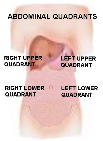
The Four abdominal Quadrants
Because of the number of the size of the abdomen and the number of organs it contains it is divided into four to facilitate diagnosis. The first line is drawn horizontally through the umbilicus, whilst the second line passes through this at right angles along the meridian line.
Diagram of the abdominal quadrants.

Diagnosis of abdominal pain
Fortunately the majority of pains in the abdomen are a result of minor conditions, an excess of acid in the stomach, wind, constipation, overeating and other minor digestive disorders. All of these are treated at home usually with over the counter medicines. When abdominal pain is more severe and is the result of an ulcer then it is treated with drugs and possibly surgery.
In the cases where doctors cannot immediately isolate or identify the cause of pain, then further tests are carried out. When this occurs the doctor will look at the patient’s medical history, and may then order x-rays, as well as blood, and urine tests. Later it may be necessary to have ultrasound tests, an endoscopy, and if necessary a CAT scan.
When to seek medical assistance
There are cases of abdominal pain, which should never be trivialised or ignored if any of the following symptoms are present then medical help should be sought.
Consult a doctor if:
- When the pain has lasted over a week.
- when the abdominal pain is accompanied by other symptoms such as vomiting, a feeling of nausea, chills, and or a fever, or whenever any of the symptoms below are present.
- Your stomach or abdomen is very tender or swollen.
- If you are either pregnant or believe that you may be pregnant.
- Eating any type of fatty food causes the pain.
- When the pain is accompanied by a vaginal discharge.
- The pain is accompanied by cystitis, or painful urination.
- The pain is accompanied by dizziness, or a shortage of breath.
You have any reason to suspect that the cause is not trivial.
Consult emergency help if:
- The onset of the pain is severe sudden and constant.
- You are vomiting blood.
- When the pain is not localised in the abdomen and there are pains in the neck shoulder and the chest.
- Either there is blood in the stool or it is a tarry black colour
Sources of the pain
- Pains in the navel area
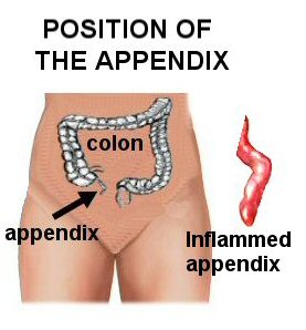
Normally when there is pain in this area it indicates intestinal disorders or the appendix. When the appendix is inflamed it fills with pus, and without treatment it can result in peritonitis, which if left untreated can kill. Symptoms are abdominal pain, nausea, vomiting, lack of appetite, a feeling of wind in the intestines, or the constant feeling of needing to have a bowel movement chills or fever.
- Pains in the epigastric area of the upper abdomen.
Persistent pain may indicate there are problems with the duodenum or small intestine, stomach, pancreas, or the gallbladder.
- Pains in the Upper right abdomen.
When the pain in this area is intense it is normally a result of an inflamed gall bladder, and it emanates out towards the centre of the abdomen and it may be felt in the back. Less commonly it may result from duodenal or pancreatic problems.
- Pains in the lower middle Abdomen.
When the pain extends to both sides of the abdomen, is often caused by colon problems. Women may be suffering from a urinary tract inflammation, or they may have an inflammatory disease of the pelvis
- Pains in the Lower left abdomen
Pain in the lower left segment of the abdomen is in the area where food is expelled from the lower colon. It may be because of an infection in the colon such as Diverticulitis, or an inflammatory bowel disease.
- Pains in the Lower right abdomen
Normally a result of problems of the colon, but appendicitis pains may spread to this region. - Referred Pain
Referred pain is when the abdominal pain has travelled along the nerve paths to cause pain in areas where there is no problem. For instance, pancreatic pain may be felt in the shoulder blades.
ARTICLE 3
SWOLLEN GLANDS
In this article
- Definition of a gland
- Position of the glands in the human body
- Endocrine and exocrine glands
- Swollen Glands
- Definition of the Lymph nodes
- Why the lymph glands swell
- Syptoms of swollen glands
- Treatment of Swollen glands
- When to seek medical advice
Definition of a Gland
The word gland comes from the Old French glandre, whose stem is the Latin glandula. In the human body a gland means two things, firstly it is a group of cells or an organ that produces a secretion. This secretion can be used in another part of the body, or they eliminate the bodies waste products. The gland synthesise the substances needed by the body, and release these for the body to utilise through ducts or they are released directly into the blood stream.
Position of the glands in the human body.

Endocrine and Exocrine glands.
Glands of the body are classified as either exocrine or endocrine types. Exocrine glands are glands that retain ducts to the body surfaces, whilst endocrine glands are isolated ductless tissue that secrete their hormones directly into the blood stream.
Endocrine and exocrine glands secrete various products, which include the hormones, enzymes, metabolites, and other molecules. In exocrine glands, the products of the glands are secreted through the duct to the surface. The endocrine glans being ductless have to secrete their products directly into the bloodstream, through capillary action.
Exocrine Glands
- Sweat Glands
- Salivary Glands
- Mammary glands
- Many of the digestive glands.
Endocrine Glands
- The word endocrine is derived from the Greek "endo," which means within, and "krine," meaning to secrete. The endocrine system is the hormonal system, they regulate the cells that have receptors for the specific hormone. The hormones are chemical messengers that influence growth, development, and metabolic activities.They include the The endocrine system includes the hypothalamus,the pituitary gland, thyroid, adrenal glands, and the gonads which are the ovaries and testes.
Swollen Glands
"Swollen gland" is a description of the lymph glands when they are enlarged, when referring to children a lymph is enlarged when it is more than one centimetre (0.4 inch) in diameter.
Definition of the Lymph nodes.
The Lymph gland are correctly called lymph nodes. The lymph nodes are small clusters of cells located along the lymphatic vessels, particularly at the neck, armpit, and groin. Their function is to filter bacteria and foreign particles from the lymph fluid. When the lymph glands have swollen the palpable nodes can be felt with the fingers in;
- Groin area the inguinal region
- Armpit the axilla region.
- There are a chain of lymph nodes on either side of the front of the neck, both sides of the neck and down past each side of the back of the neck.
- Under the jaw and chin
- Behind the ears
- On the back of the head in the occiput region
Why the Lymph Glands Swell.
Inflammatory conditions of the lymph glands normally result from infections, an abscess and cancer, there are occasionally other causes but they are very rare. As a rule of thumb the onset of minor infections are sudden and painful, being causes by infections or injury, on the other hand tumour and cancers tend to develop painlessly and very gradually. Reasons why the lymphatic glands swell include:
- An increased number of lymphocytes are present locally because they are subduing the foreign substances, known as the antigens, as they fight the infection.
- Immune reaction to an infection such as viral infections that can occur with common infections such as the cold, or more serious infections such as HIV .
- Infiltration with malignant cancer cells called metastases which have spread to the lymph glands from other organs that have cancer, or from cancers of the blood.
- Typically swollen glands cause symptoms of an upper respiratory infection, which include a runny nose, a fever, sore throat, and slight swellings under the skin in the upper part of your neck, under your chin, and possibly around the ears. It may or may be accompanied by the condition of a sore throat.
- When the lymph glands are swollen within the body they exhibit different symptoms from those underneath the skin. The blockage of the lymphs here may cause the swelling of a limb, or a chronic cough, it is also possible that the swollen gland cannot be felt externally.
- Different types of swellings are caused by different infections they may be general when there is a fungal a parasitic or a HIV infection. General swellings may accompany some of the auto immune diseases such as lupus or rheumatoid arthritis.
Treatment of Swollen Glands.
Swollen glands become inflamed when they are fighting infection, and often the subside of their own accord without treatment, within a few days. However it may take weeks for the glands to return to their normal size.
When to Seek Medical advice
Call your doctor if you have the following symptom:
- Your glands don't get smaller after two weeks
- They continue to get larger
- Your swollen glands are red and tender to the touch.
- Your glands are in a rigid position, and you cannot manipulate them with your fingertips.
- You have a fever, experience night sweating or unexplained weight loss.
- If the swelling is close to your collarbone or in the lower part of the neck
In conclusion the condition of swollen glands rarely needs emergency treatment, but they should not be ignored if there is a sign of a growing infection, or the node is so infected it needs to be surgically drained, or if the pain is severe. Children's lymphatic systems are more active, and they often feel enlarged.
ARTICLE 4
COLD FEET
In this article:
- Causes of cold feet
- Damage resulting fromcold feet
Causes of Cold Feet
The most severe cold injury is frostbite, which is true tissue freezing (ice crystals form in skin and other tissues of the body). Frostbite causes permanent damage to blood vessels and other structures. Frostnip is also ice crystal formation in tissues but only in the very outer layer of the skin. It causes no permanent damage.
Immersion injury results from exposure of wet feet (or hands) to cold temperatures at or above freezing. It develops over hours to days and damages the nerves and muscles. Like frostbite, immersion injury causes permanent damage.
Other injuries due to cold hands or feet are pernio, Raynaud syndrome, cryoglobulin formation, and cold urticaria.
![]()
ARTICLE 39
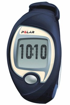
HEART RATE MONITORS
In this article
• What is a Heart rate monitor?
• The physiology of exercise.
• Relationship between V O2 max and heart rate.
• Why athlete’s recommend them
• Types Of Heart Rate Monitors.
• How to use a heart monitor
• Setting target goals for each workout
• Improving your athletic performance
• Setting realistic useful goalscatherincatherinecahterine
A heart rate monitor displays your heart rate, which is how fast it is working, or how hard it is working when you are having a workout or exercising. They are also referred to as HRM, Polar monitor, cardio monitor, and they may have an audio or visual readout. Some of the models have the bolt on goodies of a stopwatch, and they can convert how many calories the exercise is burning, whilst others can deal with preset workouts.
The physiology of exercise
Your body can be correctly identified as a combustion engine, in that it generates energy by utilizing fuel and oxygen. The cardio-vascular system delivers oxygen in the form of oxygenated blood from the lungs, to the muscles. The muscles convert this to mechanical energy, the type of energy it uses to get them working, by burning carbohydrates and fat as fuel for its engine.
The heart is no different from any other engine it can be tweaked for maximum performance. The heart can be made stronger and the muscles can become more efficient at extracting the oxygen from the blood. Within the muscle cells, the mitochondria can use their proteins in the form of enzyme systems to oxidize and burn the fuels. When you have a hard aerobic workout the aerobic system should be worked to capacity, but whilst it rests it becomes stronger. At your optimum fitness the heart is capable of pumping more blood with every beat, which means it send the oxygen needed by the muscles with less heart beats.
This level of fitness is achieved over a period of time and an accurate measurement of the rate of oxygen being "burned" by the muscles is necessary. Whilst the improvement in power is slow it is necessary to work the heart harder to achieve that level of fitness. To measure that level of fitness is also impractical outside of laboratory conditions. It would mean running on a treadmill whilst the heart rate is being monitored, and at the same time determining the concentration of oxygen in the exhaled oxygen. The disparity between the level oxygen inhaled, utilized by the muscles and then exhaled is the amount of oxygen consumed by the muscles during exercise. The rate of the oxygen consumed in liters per minute is known as V O2. When the muscles are utilizing their maximum levels of oxygen it is known as V O2 max. Whilst these tests have been carried out physiologists have discovered that there is a level beneath V O2 max in which the muscles make no gains in utilizing the oxygen.
Relationship between V O2 max and heart rate.
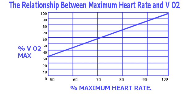
In general for most people this works out at about 55 percent of V O2max and it means that you are not overloading either your cardio vascular system or your muscular system and there will be no improvement in performance.
As the graph illustrates, 55% VO2 max corresponds to approximately 70% max heart rate.
Individual athletes have not got the equipment to determine VO2 but they can monitor their heart rate and therefore work out the percentages. The mathematics are simple as the rate is not predictable it does not have any variations according to age, sex and gender, or level of fitness.
Why athlete’s recommend them
• They precisely measure the intensity of exercise by evaluating how hard your heart is working.
• The measurement of the heart rate is the4 only effective measure of knowing how beneficial the exercise is.
• They can help to increase motivation because they are a measure of performance.
• Because you can monitor how beneficial the exercise is you can condense the exercise into the minimum amount of time necessary to get the full benefit of the exercise.
• It allows you more control over your exercise periods as you can regulate not only the length of them but also the intensity.
• It provides immediate feedback that allows you to make immediate change in the regulation of the heart rate, which means the heart, has less strain.
Types Of Heart Rate Monitors.
The majority consists of a chest transmitter that monitors the electrical signals of the heart, and then sends this rate to a receiver usually on a wristband. The “watch” type receiver shows the heart information in rates per minute.
How to use a heart monitor
The first thing you need to ascertain is your maximum heart rate, and this is normally done by an exercise stress test. If you are over the age of thirty-five then it is advisable to have a physical before undergoing this.
Setting target goals for each workout
There are different types of workouts, one is a long slow consistent workout or you have workouts using high bursts of energy. The training zone can be anywhere between seventy and ninety percent of the max heart rate, but the monitor helps you stay within the band you have set to achieve an improvement.
Improving your Athletic Performance
Working towards an improvement is the biggest single reason for using a heart rate monitor during exercise. If you are thirty years of age and you run for an hour and you cover 6.66 miles, at an average of nine minutes an hour, and your maximum heart rate is 145 a minute, what do you have to do to improve. As your fitness peak improves then running a mile will eventually not challenge the aerobic system, so to continue at the same rate will maintain fitness but not improve it. The time per mile must decrease to improve optimum fitness, or increase the workload, that is run into a head wind; although the time will be slower the heart will have to work harder.
Setting realistic useful goals.
Before improving your performance you have to decide what needs improvement, do you want to work to get faster, fitter, or stronger. Serious Training for Serious Athletes by R. Sleamaker covers plan that utilize the different target zones for different types of workout
different types of workouts. Before changing your current schedule of fitness training observe and monitor your readings for a while. This means that you are certain to know your current heart rate for a specific activity. Above all listen to your body and do not allow yourself to push your body beyond a safe limit, if in doubt, stop.
ARTICLE 40
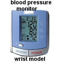
Blood pressure monitors.
In this article:
• What is the heart?
• What is blood pressure?
• How Is Blood Pressure Measured?
• What is considered a normal reading?
• Variations in blood pressure.
• What is Hypertension?
• What Type of Monitor Is Best.
What is the heart?
The heart is an incredible machine that beats approximately 100,000 times in a single day, over a lifetime that is about 2.5 billion times; but put prosaically the heart is just a pump, which sends blood through the arteries to the muscles and organs. Pumps of any description work on the principals of pressure, and put the heart under too much strain and one of the arteries can burst or the heart can stop beating. The muscles of the heart work hard, even when resting between pumps it works twice as hard as leg muscles do when sprinting.
What is Blood Pressure?
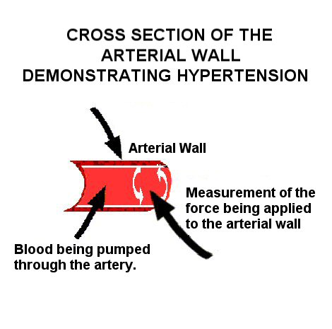
Blood pressure is the force that the blood exerts on the artery walls, as it passes through them. Blood pressure exerts two forces the first is known as systemic arterial blood pressure. Systemic pressure is when the heart pumps blood as it exits the heart through the arteries, to the circulatory system. The blood pressure from the heart in the arteries is always lower than the blood being carried back to the heart by the veins.
The secondary force is the force of the arteries when they resist the blood flow. In simplistic terms this means that blood pressure is how forcefully the pump is working per minute and how relaxed the arteries are. When the blood is forced through the arteries at too great a speed then high blood pressure or hypertension is caused
How Is Blood Pressure Measured?
Blood pressure is measured using two numbers, which are always represented as a fraction. For example if a blood pressure reading was spoken as being a hundred and ten over ninety, it would always be written as '110/90mmHg'. The mmHg means ‘millimeters of mercury’; until recently before the advent of home blood pressure monitors it was a unit of measurement measured by a sphygmomanometer, which measured your pressure over a column of mercury.
The top pressure is always the systolic pressure and the bottom is always the diastolic blood pressure. The systolic pressure is the maximum rate of heartbeat, or contraction, when it is pushing the blood out of the heart to the body. Diastolic blood pressure is the minimum pressure in the arteries between beats when the heart is relaxing and the ventricles are filling up with blood.
What is considered a normal reading?
• Isolated systolic hypertension
As we age the elasticity of the arteries alters and the blood pressure become naturally elevated. Often the diastolic resting level is normal but the systolic level starts to rise and this is called isolated systolic hypertension. Whilst it is normal it still requires medical attention and treatment.
However there is no such thing as an absolute safe standard blood, controlling blood pressure is always crucial but diabetics are more at risk and there blood pressure should be lower than most people’s.
Those who are at special risk should have a systolic pressure no pore than 140-159mmHg, and a diastolic pressure of between 90-99mmHg. In general terms anything over 160mmHg over 100mmHg is high.
Those patients with diabetes, kidney disease or have already suffered a stroke or heart attack, should aim for a lower safer pressure of 130/80 mmHg.
Variations in blood pressure
There are fairly normal variations in a blood pressure reading over a twenty-four hour period, depending on the activities you are doing or the stress you are experiencing. Blood pressure is lower whilst you sleep because this is the time you are most relaxed.
In a single day your systolic blood pressure may vary by 30 to 40 mmHg and have a proportionate increase in the diastolic, therefore it is important to measure it at the same time. If there is a large variant pressure over time it is possible to wear a cuff monitor fore a complete twenty-four hour period, this allows a doctor to attempt to isolate the cause of hypertension.
What is Hypertension?
Hypertension occurs when the blood is forced through the arteries at too high a speed. Unfortunately there are rarely any symptoms when this occurs and for this reason hypertension is known as the silent killer. Severe symptoms are headaches, confusion, coma, and sleepiness.
Prolonged untreated hypertension causes a number of life threatening complications. It increases the risks of strokes, which are blood clots or a haemorrhage in the brain. Narrowing of the arteries or atherosclerosis, a reduction in the hearts pumping mechanism and a heart attack. Other complications are an aneurysm, which is an expansion in the main artery of the chest or abdomen, which is then in danger of rupturing. It may also result in eye and renal damage.
What Type of Monitor Is Best?
A consistent monitoring is important and wrong diagnosis can result from an incorrect reading. Taking and monitoring your own blood pressure makes you feel more in control of your medical condition as you can monitor any lifestyle changes that you have made. However be aware that it may make you more nervous at least initially, and this may drive your blood pressure up. If you decide to use one the best methods is to make the measurements a part of your daily routine, like cleaning your teeth.
There are three types of blood pressure monitors, and they measure from the finger, the wrist, or the arm. The finger types are notoriously inaccurate, and should not be used. The wrist type can only produce an accurate reading if the arm is elevated to precisely the level of the heart, and as they are not user friendly they should be avoided.
The most reliable are the arm type as it gives the most accurate measurement.
.

No comments:
Post a Comment